Technische Informationen
Essential technical details of the NanoSIMS machine:
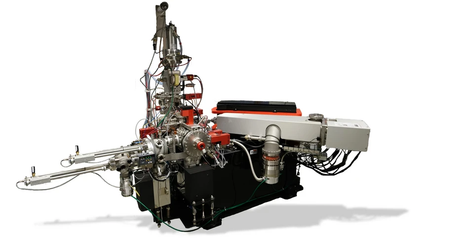
| Model | NanoSIMS 50L by CAMECA |
| Primary Ion Sources | Cs⁺ (Cesium) source and RF plasma O⁻ (Oxygen) source |
| Mass Resolution (M/ΔM) | > 10,000 (sufficient to resolve isobaric interferences) |
| Lateral Resolution | ~50 nm (depending on conditions and element), typically around 120 nm |
| Depth Resolution | < 10 nm |
| Simultaneous Detectors | Up to 7 electron multipliers (EMs) for parallel detection of isotopes/elements |
| Analyzable Elements | Most elements ranging from H to U depending on their ionization |
| Sample Types | Thin sections, polished surfaces, biological tissues, particles, etc. |
| Vacuum Requirements | Ultra-high vacuum (UHV) in analysis chamber |
| Measurement modes | Point scans, line scans, 2D raster imaging mode |
| Features | High sensitivity, high spatial resolution, multi-isotope detection |
NanoSIMS measurement modes
NanoSIMS provides several analytical modes, including imaging, line scans, and spot analysis. Each mode offers distinct advantages for investigating the elemental and isotopic composition of samples at the submicron scale.
A typical imaging measurement covers an area of 30 µm × 30 µm with 256 x 256 pixels acquired over 40 sequential image planes. This process takes about 1 hour, including acquisition time. Considering sample exchange, instrument adjustments, and data handling, we can typically analyze around 5 spots per day.
The number of sequential image planes depends on the required accuracy and sensitivity of measured ions. The relative sensivity factors depend on the ionization of different elements that can be looked up here.
To maximize instrument efficiency and accurately target regions of interest, new samples should be pre-characterized using complementary techniques prior to NanoSIMS analysis, such as light microscopy, scanning electron microscopy, transmission electron microscopy, fluorescence microscopy, or electron probe micro analysis.
Ausstattung
Besides the NanoSIMS 50L, our NanoSIMS lab is equipped with some additional instruments for sample preparation and control:
- Reflected light microscope - ZEISS Axio Imager Z2, for control of accurate sample preparation, creating maps for further SEM and NanoSIMS analyses
- Sputter coater - for coating of insulating samples with Au/Pd alloy
- Low speed saw - Allied Techcut 4, sectioning of embedded samples before final polishing
- Laminar flow box - sample mounting possible under dust free conditions
For sample mounting we can provide sample holders fitting samples ranging from filters of 5 mm diameter to up to single samples with a diameter of 2.5 cm. Beside the specific instruments for the work related to NanoSIMS, our lab runs several other analytical instruments as listed on our main homepage.
Manufacturer:
Cameca, Gennevilliers Cedex, France:
http://www.cameca.com
NanoSIMS:
http://www.cameca.com/instruments-for-research/nanosims.aspx